Sun exposure leaves a lasting impact on your skin. In Australia, where UV levels are high year-round, 40–50% of Caucasians over 40 will develop actinic keratosis. These rough, scaly patches are precancerous, meaning early detection and treatment are key to preventing progression.
At Laser Skin & Vein Clinic, we provide skin cancer checks, chemical peels, and laser resurfacing to help detect, care for, and revitalise sun-damaged skin.
With over 25 years of expertise in medical aesthetics and skin health, our highly-trained team is here to guide you through the fundamentals of actinic keratosis. Keep reading to discover its symptoms, causes, treatments and best practices for prevention.
What is actinic keratosis?
Actinic keratosis, also known as solar keratosis, is a common skin condition caused by accumulated UV damage. These pre-cancerous lesions form in the outermost layer of the skin and often present as rough, dry, or scaly patches—most commonly on areas that receive frequent sun exposure, like the face, hands, and décolletage.
While these lesions are not harmful on their own, they are a sign that your skin has experienced sun damage, and treating them early can help prevent progression into squamous cell carcinoma (SCC), a form of skin cancer.
Although the likelihood of an individual actinic keratosis developing into SCC is low—estimated at just 0.1% per lesion each year—proactive care and regular checks are essential for long-term skin health.
Where do actinic keratoses form?
Actinic keratoses most often appear on skin that is frequently exposed to sunlight, such as the face, hands, and scalp. These delicate areas, including the ears, nose, cheeks, forehead, temples, and scalp, are especially vulnerable to sun damage over time.
A specific type, known as actinic cheilitis, can also form on the lips, causing them to become persistently dry, cracked, or rough in texture.
In individuals who endure long-term sun exposure, this condition may also appear on areas beyond the face and hands, including the shoulders, chest, arms, legs, and even the tops of the feet.
Potential causes of actinic keratosis

Repeated sun exposure is the main cause of actinic keratosis, as it gradually alters the skin’s texture and appearance over time.
Whether from natural sunlight or artificial sources like tanning beds, UV rays penetrate deeply, impacting the skin’s ability to heal and regenerate.
Even subtle, daily exposure builds up over the years, contributing to skin changes that may lead to actinic keratosis. Even on overcast days, up to 80% of UV rays still reach the skin, making protection essential year-round.
This effect is intensified by reflective surfaces like sand, water, and snow, which bounce UV rays back onto the skin, increasing exposure. At higher altitudes, where the atmosphere is thinner, the sun’s impact becomes even stronger.
While the sun plays the leading role, other factors can contribute to the development of actinic keratosis:
- Environmental exposure – prolonged contact with certain chemicals, like arsenic, may increase risk.
- Radiation treatments – past exposure to ionising radiation, including X-rays, can affect skin health.
- Chronic wounds and scarring – areas of skin that struggle to heal, including burns and long-term inflammation, may become more vulnerable.
A resilient immune system helps the skin repair itself. However, if weakened—whether due to medical conditions, treatments like chemotherapy, or post-transplant immunosuppression—the skin’s ability to recover from sun exposure diminishes, increasing the likelihood of AKs forming.
Risk factors for actinic keratosis
Actinic keratosis is most common in people with prolonged sun exposure, particularly those living in tropical or subtropical climates.
Several factors can increase the likelihood of developing AK, including skin type, age, medical history, and lifestyle.
Let’s take a closer look at each risk factor:
Fair skin
People with fair skin, freckles, blonde or red hair, and blue, green, or grey eyes are at the highest risk. These skin types tend to burn rather than tan, making them more vulnerable to sun damage over time.
History of actinic keratosis or skin cancer
If you’ve had an actinic keratosis before, you’re more likely to develop another. Individuals with a history of skin cancer also face an increased risk, as these lesions can be a precursor to squamous cell carcinoma. Those previously diagnosed should have routine skin checks, typically every six months.
Family history
A genetic predisposition may play a role in developing this skin condition. Conditions like xeroderma pigmentosum, which causes extreme photosensitivity, significantly increase the risk. If close family members have experienced actinic keratoses, you may also be more susceptible.
Age and cumulative sun exposure
Actinic keratosis becomes more common with age. By the time someone reaches their 70s, they may have significantly more lesions than they did in their 50s.
Weakened immune system
Individuals with compromised immune systems—whether from conditions like HIV, post-transplant immunosuppression, or certain medical treatments—are at a higher risk. The immune system plays a crucial role in repairing sun-damaged skin, so when it’s weakened, lesions are more likely to develop.
Occupational sun exposure
People who work outdoors for extended hours, such as farmers, construction workers, and lifeguards, are at a greater risk due to chronic sun exposure. Additionally, exposure to certain industrial chemicals, like aniline dyes used in printing, may contribute to the development of actinic keratosis.
Outdoor lifestyle
Spending long periods outdoors for leisure—whether it’s sailing, fishing, golfing, or hiking—can significantly increase the likelihood of developing actinic keratosis. Without proper sun protection, repeated exposure to UV rays accelerates skin damage over time.
What does actinic keratosis look like?
Actinic keratosis can appear in different ways, but it is often characterised by rough, scaly patches that develop on sun-exposed skin. These lesions may vary in colour, texture, and severity.
Here are some common signs to look for:
- Changes in pigmentation: AKs can range from red and pink to light or dark tan, sometimes resembling freckles or age spots.
- Persistent sores: Some lesions form a crusted surface, healing and reappearing repeatedly. In rare cases, they may bleed.
- Shiny, irritated patches: Certain AKs may feel itchy, tender, or cause a pricking sensation.
- Raised pink growths: Some lesions develop a slightly elevated border and may be accompanied by tiny visible blood vessels.
Actinic keratoses can appear as isolated spots, but they are more commonly found in clusters. They are often graded based on their severity:
- Grade 1 (Mild): Slightly rough pink or grey patches with a fine scale, feeling gritty to the touch.
- Grade 2 (Moderate): Thicker, more noticeable lesions with a rough, raised texture.
- Grade 3 (Severe): Significant thickening of the skin with a pronounced, hardened surface.
- Grade 4 (Extensive): Larger affected areas spanning several centimetres, with a mix of the above characteristics.
Early detection and treatment are key to maintaining healthy, radiant skin. If you notice persistent rough patches, it’s important to have them checked by a trained healthcare professional.
How is actinic keratosis diagnosed?
A skilled professional can often recognise actinic keratosis with just a careful look and touch. These areas of sun-damaged skin have a distinct texture and appearance, making diagnosis straightforward in most cases.
If further clarity is needed, a skin biopsy may be recommended. This involves gently removing a tiny sample for lab analysis, a quick in-clinic procedure performed with a local anaesthetic for your comfort.
How to remove actinic keratoses
The best treatment for an actinic keratosis depends on several key factors, including:
- The number of lesions present
- Their location on your body
- Their appearance and severity
- Any history of skin cancer
- Underlying health conditions, such as an organ transplant
Let’s explore the different treatment options and what they involve.
Chemical peel
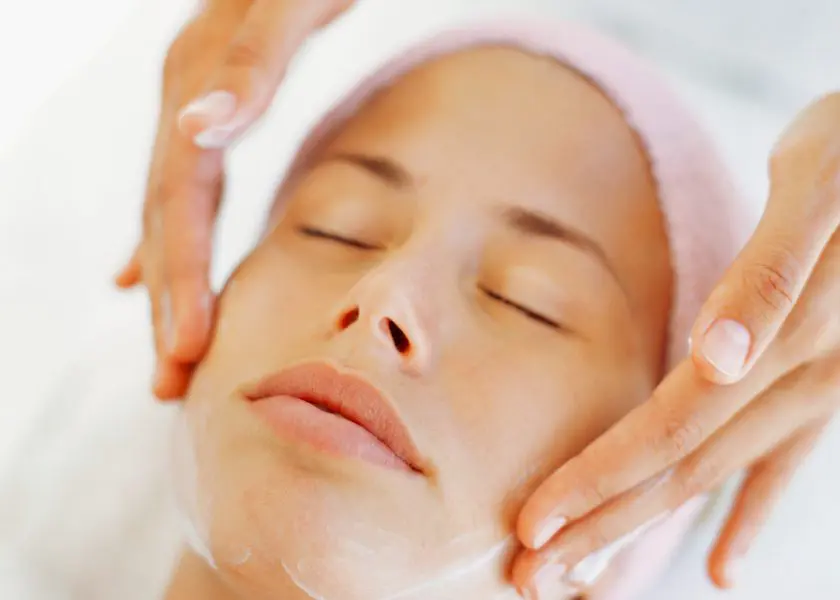
A chemical peel is a medical-grade treatment designed to remove damaged skin cells and encourage fresh, healthy skin growth.
During the procedure, a specially formulated chemical solution that targets sun-damaged areas and precancerous skin changes is applied to the affected area. This solution works by breaking down the top layers of damaged skin, prompting the body to shed them and replace them with new, healthier skin.
Afterwards, you may experience:
- Redness and swelling in the treated area
- A stinging or burning sensation as the skin reacts to the solution
- Peeling and flaking over several days as damaged skin sloughs offSmoother, healthier skin once the healing process is complete
While the healing period can be slightly uncomfortable, the long-term benefits are significant. This treatment not only helps clear up sun-damaged patches but can also improve overall skin texture and reduce the likelihood of more serious complications down the line.
Since chemical peels remove the outermost layer of skin, sun protection is essential after treatment to maintain results and prevent further damage. Your therapist may recommend specific skincare products to support healing and protect your skin moving forward.
Cryosurgery
Cryosurgery is a quick in-office procedure whereby damaged skin cells are frozen off with an ultra-cold substance such as liquid nitrogen. This extreme cold destroys abnormal cells, triggering a controlled healing response.
Some areas may require multiple treatments, especially if the damaged cells are deep or widespread.
The treated area may blister or form a scab before the damaged skin peels away, revealing new, healthier skin underneath.
Healing time depends on where the treatment is performed:
- Face: Typically heals within 5–10 days
- Hands: Recovery takes 3–4 weeks
- Legs: May take 6 weeks or longer due to slower circulation
It’s common to experience redness, swelling, crusting, or blistering at the treatment site. These are all normal signs of the skin repairing itself.
Note that for superficial skin damage, a light freeze usually leaves no lasting mark. However, deeper treatments may cause temporary or permanent lightening of the skin (hypopigmentation) or mild scarring.
Curettage
For thicker, more resilient patches of sun-damaged skin, curettage may be the most suitable treatment. This in-office procedure physically removes damaged cells, clearing the way for healthier skin to regenerate.
A specialised tool called a curet, which has a sharp, scoop-like edge, is used to carefully scrape away the damaged tissue.
In many cases, electrosurgery follows the scraping process. This involves using a pen-shaped instrument that delivers a controlled electric current to eliminate any remaining abnormal cells and prevent regrowth.
Following curettage, you may experience:
- Tenderness and redness as the area heals
- Scabbing or crusting over the treated site
- Possible changes in skin pigmentation, especially if deeper layers are affected
- A risk of scarring.
Topical medication
If you have multiple areas of sun-damaged skin, a healthcare professional may prescribe a medicated cream or gel to help remove affected cells.
This topical medication is used for superficial sun damage and offers a non-invasive alternative to in-office procedures. They work by targeting and gradually eliminating damaged skin cells over time.
During treatment, the skin may become red, inflamed, and start peeling as the damaged cells break down. A burning or stinging sensation may occur, along with scaling and crusting as the skin heals. These effects are temporary but may last several weeks, depending on the medication and individual response.
Importantly, this medication is not suitable for thicker or more resistant skin lesions. Additionally, strict sun protection is essential during and after treatment to prevent further skin damage.
Photodynamic therapy
Photodynamic therapy (PDT) is a noninvasive medical treatment used to target sun-damaged skin and certain precancerous lesions. It is particularly beneficial for individuals with multiple affected areas—such as the face and scalp—while minimising harm to surrounding healthy skin.
First, your healthcare provider applies a topical photosensitising agent, such as aminolevulinic acid (ALA) or methyl aminolevulinate (MAL), to the treatment area. This medication is left on the skin for several hours, allowing it to be absorbed by the targeted cells.
Once absorbed, the treated area is exposed to a specific light source, such as red or blue light, a pulsed-dye laser, or in some cases, controlled natural sunlight.
This activation process helps to target and destroy damaged skin cells, while aiming to preserve the surrounding skin as much as possible.
After PDT, the treated area may experience:
- Redness, swelling, and peeling as the damaged skin sheds
- Flaking or mild discomfort for a few days
- Sensitivity to sunlight—which is why avoiding direct UV exposure for at least 48 hours is crucial to prevent severe burns.
Most people require two sessions to fully treat the affected areas, with the second session scheduled about three weeks after the first.
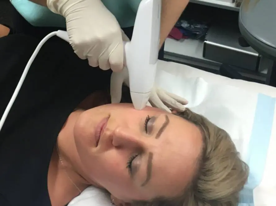
Laser resurfacing
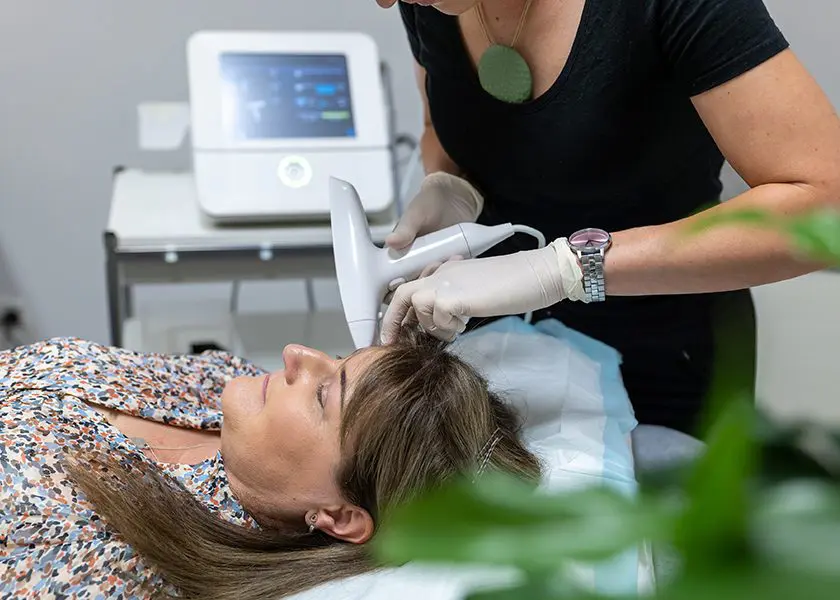
Laser resurfacing is an advanced skin treatment that is gaining popularity for removing sun-damaged skin and precancerous growths. It is often used to address actinic cheilitis, larger areas of affected skin, and persistent patches that don’t respond well to other treatments.
During the procedure, a healthcare provider uses an ablative laser device to vaporise damaged skin cells, layer by layer. This controlled removal process stimulates natural healing and promotes the growth of new, healthier skin.
Treated skin will feel raw, tender, and slightly swollen. As the area heals over the next one to two weeks, you may notice:
- Redness and peeling as damaged skin sheds
- A smoother, healthier skin surface replacing the treated area
- Mild discomfort, which can be managed with post-treatment care
- Sensitivity to sunlight
- Temporary or permanent changes in skin pigmentation
This treatment not only helps to remove precancerous skin cells but also enhances overall skin texture, reducing fine lines and uneven tone.
If you’re looking for a high-precision approach to treating sun damage while rejuvenating your skin, laser resurfacing could be an excellent choice.
How long does it take to get rid of actinic keratosis?
The time it takes for actinic keratosis to resolve depends on the size, number, and treatment approach. While some spots fade within weeks, others may take up to three months to fully clear.
Even after your skin has healed, ongoing care is key. Regular check-ups—typically once or twice a year—help maintain your skin’s health and catch any new concerns early. If your immune system is more vulnerable, more frequent visits may be recommended to ensure your skin stays its healthiest.
Tips for prevention
If you’ve experienced one of these sun-induced lesions, your skin may be more vulnerable to developing others—particularly in areas that have already been affected, such as the nose, ears, and lips.
Protecting your skin from the sun doesn’t mean avoiding the outdoors—it means embracing smart habits that help preserve its health and natural beauty.
Here’s how you can safeguard your skin every day:
- Make sunscreen part of your routine – Use a broad-spectrum SPF 30+ (or higher) every day to protect against UVA and UVB rays. For extended outdoor time, choose a water-resistant formula.
- Apply sunscreen the right way – Coat all exposed skin 10 minutes before stepping outside, and reapply every two hours—or immediately after swimming, sweating, or towelling off.
- Be sun aware – Keep an eye on the UV Index, and limit sun exposure when levels rise above 3, particularly in warmer months.
- Seek shade when the sun is strongest – Between 10 AM and 3 PM, UV exposure is at its peak, so staying in the shade can help minimise damage.
- Say no to tanning – Avoid intentional sun tanning and never use UV tanning beds.
- Dress with protection in mind – Choose lightweight, long-sleeved clothing, a broad-brimmed hat, and UV-blocking sunglasses to shield delicate areas.
- Nourish your skin from within – Nicotinamide (Vitamin B3) at 500mg twice daily may support skin health, unless otherwise advised by your healthcare provider.
- Stay in tune with your skin – Perform a full-body self-check each month, taking note of any new or changing spots.
Book a consultation for actinic keratosis treatment in Adelaide

Most actinic keratoses can be successfully treated, and early intervention helps prevent progression to skin cancer. However, sun exposure can cause new lesions to develop over time, making regular skin checks essential for ongoing protection.
At Laser Skin & Vein Clinic, we offer comprehensive skin cancer checks alongside advanced treatments like chemical peels and laser resurfacing for sun-damaged skin. With decades of experience, our team is dedicated to helping you maintain healthy, radiant skin with personalised care.
Book a consultation today and prioritise your skin’s health. We look forward to welcoming you to our Adelaide clinic.
References
- Marks R. Epidemiology of non-melanoma skin cancer and solar keratoses in Australia: a tale of self-immolation in Elysian fields. Australas J Dermatol. 1997 Jun;38 Suppl 1:S26-9. doi: 10.1111/j.1440-0960.1997.tb01004.x. PMID: 10994467.